다래(Actinidia arguta) 벌화분의 페놀아마이드 및 지방산 유도체의 분리 및 구조 동정
Abstract
Bee pollen of Actinidia arguta (A. arguta) has been recognized as a functional material containing diverse bioactive compounds. however, previous studies have primarily focused on extract-level biological activities, and information on individual active constituents remains limited. In this study, major compounds were isolated from A. arguta bee pollen, and their structures were elucidated using chromatographic techniques in combination with LC-MS and NMR analyses. As a result, five compounds were identified, including three phenolamide compounds belonging to dicoumaroylputrescine and coumaroyl spermidine derivatives, as well as terrestribisamide and roughanic acid. The phenolamide compounds were characterized by molecular ion peaks in mass spectrometry, and their structures were further supported by characteristic fragmentation patterns. In addition, geometric isomers of coumaroyl spermidine derivatives were distinguished based on differences in coupling constants of olefinic protons observed in proton nuclear magnetic resonance spectra. Notably, terrestribisamide was confirmed as a methoxylated derivative of dicoumaroylputrescine, indicating structural diversity of phenolamide compounds in A. arguta bee pollen. The presence of roughanic acid further suggested that the chemical composition of bee pollen includes not only phenolic compounds but also fatty acid-derived metabolites. These results provide chemical evidence for the structural diversity of major constituents in A. arguta bee pollen and are expected to serve as fundamental data for future studies on biological activities and the development of functional materials.
Keywords:
Actinidia arguta, Bee pollen, Dicoumaroylputrescine, Coumaroyl spermidine derivatives, Terrestribisamide, Roughanic acid서 론
벌화분(bee pollen)은 꿀벌이 식물의 화분을 수집하여 화밀 및 효소와 함께 응집시킨 천연물로, 단백질, 아미노산, 지방산, 비타민 및 무기질 등 다양한 영양성분을 함유하고 있어 기능성 식품 소재로 주목받고 있다(Rodriguez Flores et al., 2024; Gambaro et al., 2025; Romero Villarreal et al., 2026). 특히 벌화분에는 페놀화합물, 플라보노이드와 같은 다양한 생리활성 물질이 존재하며, 이들은 항산화, 항염증 및 항암 활성과 밀접하게 관련된 것으로 보고되고 있다(Braciuniene 2026; Mengesha et al., 2026; Sanlier et al., 2026).
최근 연구에서는 벌화분의 생리활성이 총 페놀 함량보다 특정 구조를 갖는 개별 화합물에 의해 결정되는 것으로 보고되고 있다(Li et al., 2026; Liu et al., 2026). 선행 연구를 통해서 다래(Actinidia arguta) 벌화분 추출물은 전립선 암세포의 증식 억제 효과뿐만 아니라 항염 활성 등 다양한 생리활성을 나타내는 것으로 확인되었다(Kim et al., 2024; Lee et al., 2023). 특히 이러한 생리활성은 벌화분에 함유된 2차 대사산물이 기인하는 것으로 알려져 있으며, 이들 화합물은 활성산소 제거, 염증 매개인자 억제 및 세포 신호전달 조절 등을 통해 기능성을 발현하는 것으로 보고되고 있다(Pieracci et al., 2026; Saker et al., 2026).
지금까지의 벌화분 연구는 대부분 추출물 수준에서의 생리활성 평가에 집중되어 있으며, 개별 화합물의 분리 및 구조 규명에 관한 연구는 제한적으로 수행되어 왔다(Izo et al., 2025; Liu et al., 2025). 또한 벌화분은 식물 기원에 따라 화학적 조성이 크게 달라지므로 특정 밀원 유래 벌화분에 대한 심층적인 성분 분석이 필요하다(Joo et al., 2025; Stebuliauskaite et al., 2025; Yildirim and Gercek, 2025). 따라서 본 연구에서는 다래 벌화분으로부터 화학적 성분을 분리하고, LC-MS 및 NMR 분석을 통해 구조를 규명하여 다래 벌화분의 화학적 특성과 생리활성 물질 기반을 구축하고자 하였다.
실험 및 방법
1. 실험재료
본 연구에 사용된 다래 벌화분은 2024년 청양군의 농가에서 생산한 것으로 구입하였다. 구입 후 벌화분은 수분 제거를 위해 동결건조하였다.
실험에 사용된 메탄올, n-hexane, ethyl acetate 등 유기용매는 HPLC grade 시약을 사용하였으며, silica gel (70~230 mesh, Merck, Germany)과 Sephadex LH-20 (GE Healthcare, Sweden)은 column chromatography에 사용하였다.
2. 추출 및 분획
건조된 다래 벌화분 1 kg을 메탄올 3 L를 사용하여 3회 반복 추출하였다. 각 추출액을 합쳐 감압농축하여 메탄올 추출물 268.87 g을 얻었다.
농축된 추출물에 증류수를 첨가하여 현탁시킨 후, n-hexane 및 ethyl acetate를 이용하여 순차적으로 용매 분획을 수행하였다. 그 결과 n-hexane 분획 64.96 g과 ethyl acetate 분획 16.07 g을 얻었으며, 각 분획은 이후 분리 및 정제에 사용하였다.
3. 분리 및 정제
각 분획은 TLC 및 HPLC 분석을 통해 성분 패턴을 확인하였으며, 유사한 패턴을 나타내는 분획을 통합하여 분리 과정에 사용하였다. n-hexane 분획과 ethyl acetate 분획을 혼합한 후 silica gel open column chromatography (CC, 5×80 cm)를 수행하였다. 이동상으로는 n-hexane : acetone 혼합 용매를 사용하여 gradient 조건으로 용리하였다. 용리 조건은 Table 1에 나타내었다.

Elution conditions for open column chromatography
그 결과 총 8개의 분획(1~8)을 얻었다. 각 소분획은 TLC 및 HPLC 분석을 통해 주요 성분을 확인한 후, 반복적인 column chromatography 및 HPLC를 통해 단일 화합물로 분리하였다.
분획 3은 주요 화합물을 포함하는 것으로 확인되어 silica gel column chromatography를 통해 재분리하였으며, 반복적인 정제를 통해 단일 화합물을 획득하였다. 또한 분획 7은 Sephadex LH-20 column chromatography 및 preparative HPLC를 이용하여 추가 정제하였으며, 최종적으로 순수 화합물을 분리하였다.
4. 구조동정
분리된 화합물의 구조 규명을 위해 질량분석(MS) 및 핵자기공명분석(NMR)을 수행하였다. NMR 분석은 1H-NMR, 13C-NMR을 이용하여 수행하여 화합물의 구조를 해석하였다.
결과 및 고찰
다래 벌화분 추출물로부터 다양한 액체 크로마토그래피 기법을 이용하여 성분을 분리하였다. 그 결과, 총 5가지 화합물이 분리·동정되었다. 각 화합물의 구조는 LC-MS 및 NMR 분석을 통해 규명되었다. 분리된 각 화합물에 대한 정보는 Table 2에 정리하였다.
Structural chracterization of compounds isolated from Actinidia arguta pollen
1. Roughanic acid의 분리 및 구조 동정
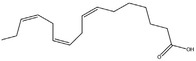
분획 3 (1.397 g)은 silica gel column (CC, 3×60 cm)을 이용하여 n-hexane : acetone 및 methanol을 포함한 용매 조건에서 분리하였으며, 그 결과 분획 3-1부터 3-4까지의 소분획을 얻었다. 이 중 분획 3-2 (891.5 mg)를 추가로 정제하였다. 정제된 화합물(520 mg)은 HE-3 분획으로부터 분리된 화합물이다. 구조는 LC-MS 및 NMR 분석을 통해 규명하였다. LC-MS 분석 결과, 본 화합물은 m/z 250.19의 주요 피크를 나타내었으며, 이는 분자량 250.38 g/mol에 해당하는 화합물의 [M - H]- 이온으로 판단된다. 또한 MS/MS 분석에서 관찰된 fragment ion은 불포화 지방산에서 흔히 나타나는 탄소 사슬 절단에 의한 특징적인 fragmentation pattern과 일치하였다(Ares et al., 2018; Yoo and Chung, 2025; Turkol et al., 2026). Fig. 1의 1H-NMR 스펙트럼에서는 δ 5.3~5.5 ppm 영역에서 olefinic proton 신호가 관찰되어 이중결합의 존재를 확인할 수 있었으며, δ 2.0~2.3 ppm 영역에서는 allylic methylene proton 신호가 확인되었다. 또한 δ 1.2~1.5 ppm 영역에서는 긴 aliphatic chain에 해당하는 methylene proton 신호가 넓은 피크로 나타났으며, δ 0.8~0.9 ppm에서는 말단 methyl proton 신호가 관찰되었다. 이러한 신호 패턴은 불포화 지방산에서 일반적으로 보고되는 특징과 일치한다. Fig. 2의 13C-NMR 스펙트럼에서는 δ 175~180 ppm 영역에서 carboxylic acid에 해당하는 carbonyl carbon 신호가 확인되었고, δ 128~132 ppm 영역에서는 olefinic carbon 신호가 관찰되었다. 또한 δ 22~35 ppm 영역에서는 aliphatic methylene carbon 신호들이 다수 확인되었으며, δ 14 ppm 부근에서 terminal methyl carbon 신호가 나타났다. 이러한 결과는 식물 유래 지방산의 전형적인 분광학적 특성과 잘 일치한다. 이상의 LC-MS 및 NMR 분석 결과는 기존 문헌에 보고된 roughanic acid의 분광학적 데이터와 부합하였으며, 따라서 본 화합물은 roughanic acid로 동정하였다(Al Qudah and Abu Zarga, 2010).
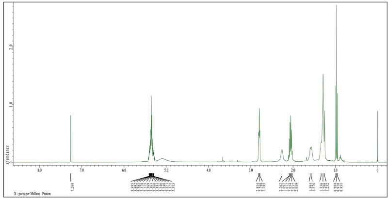
1H-NMR spectrum of roughanic acid (600 MHz, CDCl3).
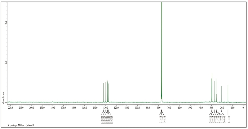
13C-NMR spectrum of roughanic acid (150 MHz, CDCl3).
2. N,Nʹ-(E)-dicoumaroylputrescine의 분리 및 구조 동정
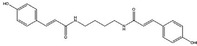
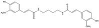
분획 7에 대하여 CHCl3와 MeOH를 용매로 실리카겔 오픈 컬럼 크로마토그래피를 수행한 결과, 총 9개의 소분획물을 얻었다. 소분획물 중 양이 많은 분획 7-7을 sephadex로 오픈 컬럼 크로마토그래피하여 4개 분획물을 얻고, 그중 분획 7-7-2를 HPLC로 물질 분리를 계속하여 분획 7-7-2-6을 분리하였다. 분리된 화합물(16 mg)의 LC-MS 분석 결과, m/z 380.17의 주요 피크가 관찰되었으며, 이는 분자량 382.45 g/mol에 해당하는 화합물의 [M - H]- 이온으로 판단된다. 또한 MS/MS 분석에서 m/z 189 및 191의 fragment ion이 관찰되었는데, 이는 p-coumaroyl 구조에서 유래하는 특징적인 fragment로 보고된 바 있다. Fig. 3의 1H-NMR 스펙트럼에서는 δ 6.2~7.5 ppm 영역에서 방향족 proton 신호와 함께 trans-olefinic proton 신호가 관찰되었으며, 특히 큰 coupling constant (J≈15~16 Hz)를 갖는 이중결합 신호는 p-coumaroyl기의 trans 형태를 명확히 지지한다. 또한 δ 3.0~3.5 ppm 영역에서는 putrescine 골격에 해당하는 methylene proton 신호가 관찰되었으며, 이는 polyamine 구조의 존재를 나타낸다. Fig. 4의 13C-NMR 스펙트럼에서는 δ 165~170 ppm 영역에서 amide carbonyl carbon 신호가 확인되었고, δ 115~145 ppm 영역에서는 방향족 및 olefinic carbon 신호가 관찰되었다. 또한 δ 35~45 ppm 영역에서는 polyamine chain에 해당하는 methylene carbon 신호가 확인되었다. 이러한 MS 및 NMR 분석 결과는 두 개의 p-coumaroyl기가 putrescine 골격에 결합된 phenolamide 구조와 잘 일치하며, 기존에 보고된 N,Nʹ-dicoumaroylputrescine의 분광학적 데이터와 부합하였다(Choi et al., 2007).
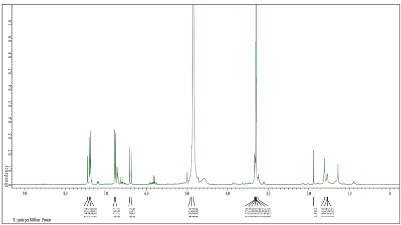
1H-NMR spectrum of N,Nʹ-dicoumaroylputrescine (600 MHz, CDCl3).
13C-NMR spectrum of N,Nʹ-dicoumaroylputrescine (150 MHz, CDCl3).
3. N1(E),N5(Z),N10(E)-tri-p-coumaroyl spermidine의 분리 및 구조 동정
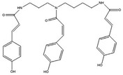
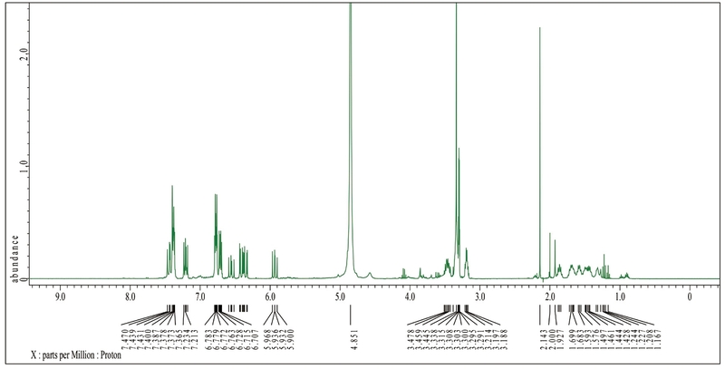
분획 7-7-2를 HPLC 분리를 수행하여 추가적인 단일 화합물 7-7-2-8을 분리하였다. 해당 화합물의 구조는 MS 및 NMR 스펙트럼 데이터를 종합적으로 분석하여 규명하였다. MS 분석에서 주요 분자 이온 피크는 m/z 582 [M - H]-로 확인되었으며, 이는 spermidine 골격에 세 개의 p-coumaroyl기가 결합된 구조에 부합하는 분자량을 나타낸다. 또한 MS/MS 분석에서 관찰된 주요 fragment ion들은 coumaroyl moiety의 순차적 탈리 패턴을 보여 해당 구조를 지지하였다. Fig. 5의 1H-NMR(600 MHz, CDCl3) 스펙트럼에서는 δ 7.40~7.60 ppm 영역에서 aromatic proton 신호가 관찰되었으며, δ 6.20~6.50 ppm 부근에서 olefinic proton 신호가 나타났다. 특히, δ≈7.5 ppm (d, J≈15~16 Hz) 및 δ≈6.3 ppm (d, J≈15~16 Hz)의 coupling constant는 trans (E) 이중결합의 존재를 명확히 나타낸다. 반면 일부 olefinic proton에서 상대적으로 작은 coupling constant가 관찰되어 Z configuration이 일부 포함됨을 나타낸다. 또한 δ 3.0~3.5 ppm 영역에서는 spermidine 골격의 methylene proton 신호가 확인되었으며, 이는 아민 사슬 구조의 존재를 뒷받침한다. Fig. 6의 13C-NMR (150 MHz, CDCl3) 스펙트럼에서는 δ 165~170 ppm 영역에서 amide carbonyl carbon 신호가 관찰되었고, δ 140~150 ppm에서는 aromatic 및 olefinic carbon 신호가 나타났다. δ 115~130 ppm 영역의 다수의 피크는 p-coumaroyl 고리 구조에 해당하며, δ 30~50 ppm 영역에서는 spermidine 사슬의 aliphatic carbon 신호가 확인되었다. 이와 같은 MS 및 NMR 데이터를 종합적으로 비교 분석한 결과, 기존 문헌에 보고된 N1(E),N5(Z),N10(E)-tri-p-coumaroyl spermidine의 스펙트럼 데이터와 일치하였으며, 최종적으로 해당 구조로 동정하였다(Ma et al., 2001).
1H-NMR spectrum of N1(E),N5(Z),N10(E)-tri-p-coumaroyl spermidine (600 MHz, CDCl3).
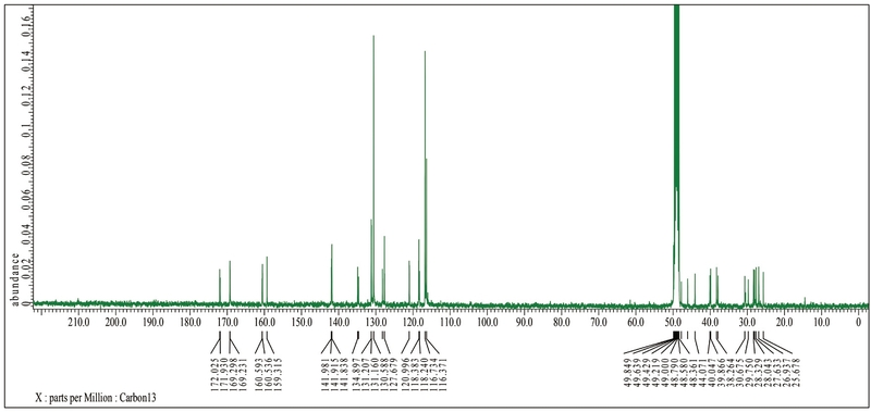
13C-NMR spectrum of N1(E),N5(Z),N10(E)-tri-p-coumaroyl spermidine (150 MHz, CDCl3).
4. N1,N5,N10-(E)-tri-p-coumaroylspermidine의 분리 및 구조 동정
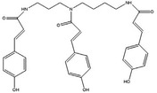
분획 7-7-2를 반복적인 preparative HPLC 분리를 수행하여 추가적인 화합물 7-7-2-9 (28 mg)를 분리하였다. ESI-MS 분석에서 주요 분자 이온 피크는 m/z 582 [M - H]-로 확인되었으며, 이는 spermidine 골격에 세 개의 p-coumaroyl기가 결합된 구조와 일치한다. MS/MS 분석에서도 coumaroyl기의 탈리에 의한 특징적인 fragment ion 패턴이 관찰되었다. Fig. 7의 1H-NMR (600 MHz, CDCl3) 스펙트럼에서 δ 7.40~7.60 ppm 영역에 aromatic proton 신호가 나타났으며, δ 6.20~6.50 ppm 영역에서 olefinic proton 신호가 관찰되었다. 특히 모든 olefinic proton에서 약 J≈15~16 Hz의 큰 coupling constant가 확인되어 세 개의 p-coumaroyl의 이중결합이 모두 trans (E) configuration임을 명확히 나타낸다. 또한 δ 3.0~3.5 ppm 영역에서는 spermidine 사슬에 해당하는 methylene proton 신호가 관찰되었으며, 이는 아민 골격 구조를 지지한다. Fig. 8의 13C-NMR (150 MHz, CDCl3) 스펙트럼에서는 δ 165~170 ppm에서 amide carbonyl carbon 신호가 확인되었고, δ 140~150 ppm에서는 aromatic 및 olefinic carbon 신호가 나타났다. δ 115~130 ppm 영역의 신호는 p-coumaroyl 고리 구조에 해당하며, δ 30~50 ppm 영역에서는 spermidine 골격의 aliphatic carbon 신호가 확인되었다. 특히, 동일한 분자량(m/z 582)을 갖는 화합물 7-7-2-8과 비교하여 olefinic proton의 coupling constant 차이를 통해 이성질체임이 확인되었다(Li et al., 2013).
1H-NMR spectrum of N1,N5,N10-(E)-tri-p-coumaroylspermidine (600 MHz, CDCl3).
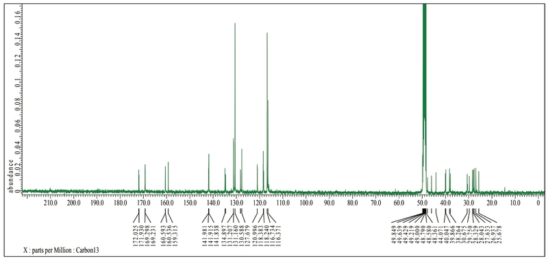
13C-NMR spectrum of N1,N5,N10-(E)-tri-p-coumaroylspermidine (150 MHz, CDCl3).
5. Terrestribisamide의 분리 및 구조 동정
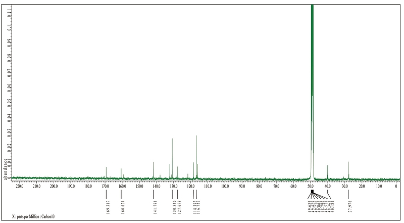
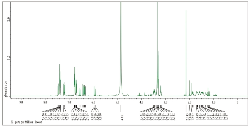
분획 7-9를 HPLC 크로마토그램에 따라 분획하여 총 9개의 소분획물을 얻었으며, 그중 7-9-3을 정제 과정을 통해 단일 화합물로 확보하였다. 해당 화합물(28 mg)은 ESI-MS 분석에서 분자 이온 피크는 m/z 396 [M - H]-로 확인되었으며, 이는 dicoumaroyl putrescine 유도체에 methoxy기가 도입된 구조와 일치하였다. 또한 MS/MS 분석에서 coumaroyl moiety의 특징적인 fragment ion이 관찰되어 해당 구조를 지지하였다. Fig. 9의 1H-NMR (600 MHz, CDCl3) 스펙트럼에서는 δ 7.0~7.6 ppm 영역에서 aromatic proton 신호가 관찰되었고, δ 6.2~6.5 ppm 부근에서 olefinic proton 신호가 확인되었다. 특히 J≈15~16 Hz의 coupling constant를 나타내는 신호는 trans(E) configuration의 존재를 시사하며, 이는 hydroxycinnamic acid 유도체에서 일반적으로 관찰되는 특징이다. 또한 δ 3.7~3.9 ppm 영역에서 methoxy (-OCH3) proton 신호가 추가적으로 관찰되어 기존의 dicoumaroyl putrescine 구조와 차별되는 특징을 나타냈으며, δ 2.5~3.5 ppm 영역에서는 putrescine 사슬에 해당하는 methylene proton 신호가 확인되었다. Fig. 10의 13C-NMR (150 MHz, CDCl3) 스펙트럼에서는 δ 165~170 ppm 영역에서 amide carbonyl carbon 신호가 관찰되었으며, δ 145~150 ppm 및 115~130 ppm 영역에서 aromatic 및 olefinic carbon 신호가 확인되었다. 특히 δ 55~60 ppm 영역에서 methoxy carbon 신호가 나타나 methoxylation 구조를 뒷받침하였다.
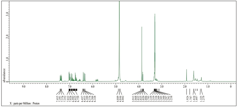
1H-NMR spectrum of terrestribisamide (600 MHz, CDCl3).
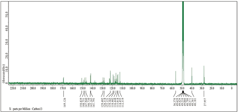
13C-NMR spectrum of terrestribisamide (150 MHz, CDCl3).
이와 같은 MS 및 NMR 데이터를 종합적으로 분석한 결과, 7-9-3 화합물은 기존에 보고된 N,Nʹ-(E)-dicoumaroyl putrescine의 methoxylated 유도체인 terrestribisamide로 동정되었다. 특히, 본 화합물은 앞서 분리된 N,Nʹ-(E)-dicoumaroyl putrescine과 비교하여 methoxy기가 추가된 구조적 변형을 나타내며, 이는 벌화분 유래 phenolic amide 화합물의 구조 다양성을 시사한다(Farhat et al., 2025).
결 론
본 연구는 다래 벌화분에서 주요 화합물을 분리하고, LC-MS 및 NMR 분석을 기반으로 이들의 화학적 구조를 규명하였다. 그 결과, dicoumaroylputrescine과 tri-p-coumaroylspermidine 계열 화합물 3종을 포함하여 terrestribisamide 및 roughanic acid 등 총 5종의 화합물을 성공적으로 분리·동정하였다. 특히 spermidine 및 putrescine 골격에 hydroxycinnamic acid 유도체가 결합된 페놀아마이드 화합물은 다래 벌화분의 주요 구성 성분으로 확인되었으며, 이 중 coumaroylspermidine 계열 화합물은 동일한 분자량을 가지면서도 기하 이성질체로 존재함을 확인하였다. 이러한 이성질체는 수소 핵자기공명 분석에서 올레핀 수소의 결합상수 차이를 통해 구분되었다. 또한 terrestribisamide는 dicoumaroylputrescine의 메톡실화 유도체로 확인되어, 다래 벌화분 내 페놀아마이드 화합물의 구조적 다양성을 보여주었다. 한편 roughanic acid의 존재는 벌화분이 페놀성 화합물뿐만 아니라 지방산 유래 대사산물도 함께 포함하는 복합적인 화학적 조성을 지님을 시사한다. 다양한 연구를 통해서 밝혀진 Phenolamide 계열 화합물이 항산화, 항염 및 효소 저해 활성과 같은 생리활성을 나타내는 점을 고려할 때, Actinidia arguta 벌화분으로부터 확인된 phenolamide 화합물은 벌화분의 항산화 활성 및 건강 기능성과 밀접한 관련이 있을 것으로 판단된다. 본 연구 결과는 다래 벌화분의 주요 화합물을 개별 성분 수준에서 분리하고 구조를 규명함으로써, 기존의 추출물 중심 생리활성 연구를 보완하는 화학적 근거를 제시하였다. 이는 향후 다래 벌화분의 생리활성 기전 규명 및 기능성 소재로서의 활용 가능성을 평가하는 데 중요한 기초 자료로 활용될 수 있을 것으로 판단된다.
Acknowledgments
본 연구는 농촌진흥청 국립농업과학원의 과제 RS-2024-00332514 지원을 받아 수행되었습니다.
References
-
Al Qudah, M. A. and M. H. Abu Zarga. 2010. Chemical constituents of Sisymbrium irio L from Jordan. Nat. Prod. Res. 24: 448-456.
[https://doi.org/10.1080/14786410903388025]

-
Ares, A. M., M. J. Nozal and J. Bernal. 2018. Extraction, chemical characterization and biological activity determination of bee pollen. J. Pharm. Biomed. Anal. 147: 110-124.
[https://doi.org/10.1016/j.jpba.2017.08.009]
-
Braciuniene, A., R. Strebliukaite, M. Liaudanskas, Z. Viksnaite, N. Sutkeviciene and S. Trumbeckaite. 2026. Changes in the amino acid composition of bee collected pollen during 15 months of storage in fresh frozen and dried forms. Foods 15: 207.
[https://doi.org/10.3390/foods15020207]
-
Choi, S. W., Y. S. Kwon, S. K. Kim, G. H. Kim and K.S. Kim. 2007. Antioxidant and antimelanogenic activities of polyamine conjugates from corn bran and related hydroxycinnamic acids. J. Agric. Food Chem. 55: 3920-3925.
[https://doi.org/10.1021/jf0635154]
-
Farhat, H., B. Bouzayani, A. Ammar, N. Tene, I. Hadrich, M. Treilhou, R. Mezghani, M. Trigui and M. Siala. 2025. Mass spectrometry based metabolomic analysis and biological activities of alkaloids from Haloxylon persicum Bunge leaves extracts. Chem. Afr. 8: 4355-4372.
[https://doi.org/10.1007/s42250-025-01482-y]
-
Gambaro, A., M. Miraballes, N. Urruzola, M. Kniazev, C. Dauber, M. Romero, A. M. Fernandez Fernandez, A. Medrano, E. Santos and I. Viezzi. 2025. Physicochemical composition and bioactive properties of Uruguayan bee pollen from different botanical sources. Foods 14: 1689.
[https://doi.org/10.3390/foods14101689]
-
Izo, E., M. Turhan, Y. Abdullah, C. Caglayan and I. Gulcin. 2025. Determination of antioxidant antidiabetic anticholinergic antiglaucoma properties and comprehensive phytochemical content by LC MS MS of Bingol honeybee pollen. Food Sci. Nutr. 13: 4531.
[https://doi.org/10.1002/fsn3.4531]
-
Joo, Y., E. Shin, H. Kim, M. K. Lee and S. B. Kim. 2025. Molecular networking guided annotation of flavonoid glycosides from Quercus mongolica bee pollen. Int. J. Mol. Sci. 26: 7930.
[https://doi.org/10.3390/ijms26167930]
-
Kim, S. K., M. S. Lee, S. I. Moon, H. Y. Kim, H. M. Choi and S. O. Woo. 2024. Inhibition of inflammation response and the immune related protein by treatment of Actinidia arguta pollen in Raw264.7 macrophage. J. Apic. 39: 223-231.
[https://doi.org/10.17519/apiculture.2024.09.39.3.223]
-
Lee, H. J., S. J. Kim, H. J. Han, K. H. Kim, S. H. Kim, S. K. Kim, H. M. Choi, H. J. Moon, S. I. Lee and S. O. Woo. 2023. Inhibitory effect of the darae pollen Actinidia arguta extract on the growth of LNCaP human prostate cancer cell line. J. Apic. 38: 77-82.
[https://doi.org/10.17519/apiculture.2023.04.38.1.77]
-
Li, K., J. Zhao, R. Wang, J. Cao, Q. Liu, W. Ji, F. Luo, J. Liu, X. Xu and L. Luo. 2026. Bioaccessibility of phenolics and anti inflammatory effects during in vitro simulated digestion of enzymatically broken bee collected pollen. Food Chem. 508: 148318.
[https://doi.org/10.1016/j.foodchem.2026.148318]
-
Li, W. C., X. Y. Wang, P. C. Lin and N. Hu. 2013. Preparative separation and purification of four cis-trans isomers of coumaroylspermidine analogs from safflower by high-speed counter-current chromatography. J. Chromatogr. B 938: 75-79.
[https://doi.org/10.1016/j.jchromb.2013.08.012]
-
Liu, L., Z. Sun, A. Liang, R. Zhang, S. He, Y. He, M. Zhang, X. Li and X. Wu. 2025. Screening of characteristic metabolites in bee pollen from different floral sources based on high resolution mass spectrometry. Foods 14: 4305.
[https://doi.org/10.3390/foods14244305]
-
Liu, W., R. Liu, Y. Han, X. Chen and Q. Lu. 2026. Phenolamide extract of apricot bee pollen alleviates DSS induced ulcerative colitis in mice by reducing oxidative stress modulating inflammation and regulating gut microbiota. Antioxidants 15: 403.
[https://doi.org/10.3390/antiox15030403]
-
Ma, C. M., N. Nakamura and M. Hattori. 2001. Inhibitory effects on HIV-1 protease of tri-p-coumaroylspermidine from Artemisia caruifolia and related amides. Chem. Pharm. Bull. 49: 915-917.
[https://doi.org/10.1248/cpb.49.915]
-
Mengesha, A. A., S. H. Mekuria and H. W. Woldemichael. 2026. Recent trends in postharvest handling preservation processing and application of bee pollen. Discov. Food
[https://doi.org/10.1007/s44187-026-00908-x]
-
Pieracci, Y., B. D’Ambrosio, G. Filannino, T. Lombardi and L. Pistelli. 2026. Volatilome and nutraceutical composition of bee pollen may serve as indicators of seasonal and botanical origin. Horticulturae 12: 376.
[https://doi.org/10.3390/horticulturae12030376]
- Rodriguez Flores, M. S., Y. Saker, M. C. Seijo, S. Harbane and O. Escuredo. 2024. Functional antioxidant assessment of bee pollen based on phenolic composition botanical origin and composite index validation. Appl. Sci. 14: 2674.
-
Romero Villarreal, K. M., I. Lobo Fafard, M. A. Alcala Orozco, J. Carrascal, B. J. Anaya, D. F. Tirado and D. C. Mantilla Escalante. 2026. Biotechnological transformation of tropical bee pollen through enzymatic and bacterial processes impact on composition and antioxidant activity. Appl. Sci. 16: 1471.
[https://doi.org/10.3390/app16031471]
-
Saker, Y., O. Escuredo, M. C. Seijo, S. Harbane, A. Oulehadj and M. S. Rodriguez Flores. 2026. Bioactive properties of Algerian bee pollen influence of botanical origin on polyphenol content and antioxidant capacity. Foods 15: 202.
[https://doi.org/10.3390/foods15020202]
-
Sanlier, N., E. F. Kaya and I. Yucel. 2026. Health effects of bee products a comprehensive review. Food Sci. Nutr. 14: 71165.
[https://doi.org/10.1002/fsn3.71165]
-
Stebuliauskaite, R., M. Liaudanskas, V. Zvikas, V. Ceksteryte, N. Sutkeviciene, S. Sorkyte, A. Braciuliene and S. Trumbeckaite. 2025. Changes in ascorbic acid phenolic compound content and antioxidant activity in vitro in bee pollen depending on storage conditions impact of drying and freezing. Antioxidants 14: 462.
[https://doi.org/10.3390/antiox14040462]
-
Turkol, M., C. Y. Mavis and S. Yilmis. 2026. From chemistry to functionality HPLC DAD LC MS MS characterization of bee product enriched Prunus spinosa L kombucha with in vitro antidiabetic activity and bioaccessibility. ACS Omega 11: 14048-14063.
[https://doi.org/10.1021/acsomega.6c00008]
-
Yildirim, N. and Y. C. Gercek. 2025. Preliminary characterization of monofloral Helianthus annuus L bee pollen using advanced analytical techniques a comparative study with polyfloral pollen. Food Sci. Nutr. 13: 71016.
[https://doi.org/10.1002/fsn3.71016]
-
Yoo, S. E. and H. Y. Chung. 2025. Quality properties of acorn bee pollen extracts. Prev. Nutr. Food Sci. 30: 591-598.
[https://doi.org/10.3746/pnf.2025.30.6.591]